Biology, 31.07.2019 02:30 xXCoryxKenshinXx
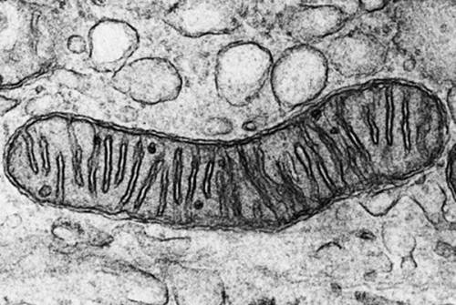
Apathologist who wants to examine a patient's liver cells to determine if the mitochondria have an internal structural defect will likely need to use a a pathologist who wants to examine a patient's liver cells to determine if the mitochondria have an internal structural defect will likely need to use a transmission electron microscope. scanning electron microscope. chromatin stain. light microscope.

Answers: 1
Another question on Biology
Biology, 22.06.2019 02:30
Are the earth's ocean floor and continents the same age geologically? why or why not?
Answers: 1
Biology, 22.06.2019 03:30
Cold case files recently began re-investigating an old murder case. the murder took place in the park; a young man, james, was hit over the head with a brick and killed. police suspected the jealous former boyfriend of james' wife, karen. whoever killed james may have washed their hands in a near-by bird bath as detectives found blood in the bird bath. since the murder took place before dna fingerprinting was available, the suspected killer, ronnie, went free. detectives are now reviewing the blood evidence using dna fingerprinting. based on the dna fingerprints of all possible suspects, who is james' killer?
Answers: 2
Biology, 22.06.2019 04:00
Will mark brainliest i only need the ! 1.use ten beads and a centromere of one color to construct the long chromosome. use ten beads and a centromere of a second color to construct the second chromosome in the long pair. make a drawing of the chromosomes in the space below. 2. for the second pair of chromosomes, use only five beads. 3. now model the replication of the chromosomes. make a drawing of your model in the space below. part b: meiosis i during meiosis i, the cell divides into two diploid daughter cells. 4. pair up the chromosomes to form tetrads. use the longer tetrad to model crossing-over. make a drawing of the tetrads in the space below. 5. line up the tetrads across the center of your “cell.” then model what happens to the chromosomes during anaphase i. 6. divide the cell into two daughter cells. use the space below to make a drawing of the result. part c: meiosis ii during meiosis ii, the daughter cells divide again. 7. line up the chromosomes at the center of the first cell, one above the other. separate the chromatids in each chromosome and move them to opposite sides of the cell. 8. repeat step 7 for the second cell. 9. divide each cell into two daughter cells. use the space below to make a drawing of the four haploid cells
Answers: 1
Biology, 22.06.2019 11:30
Which organism can most likely be classifild as domain bateria
Answers: 1
You know the right answer?
Apathologist who wants to examine a patient's liver cells to determine if the mitochondria have an i...
Questions


Mathematics, 21.04.2020 16:57


History, 21.04.2020 16:57

Computers and Technology, 21.04.2020 16:57


Mathematics, 21.04.2020 16:57
Physics, 21.04.2020 16:57

History, 21.04.2020 16:57
Mathematics, 21.04.2020 16:57