Biology, 11.11.2020 06:00 zachthomas024
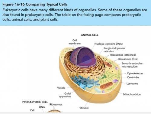
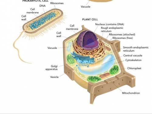
Using Figure 16-16 on the next page as a guide, draw your own models of a prokaryotic cell, a plant cell, and an animal cell. Then use each of the vocabulary words from this lesson to label your cells. Describe any differences between the models in Figure 16-16 and your models. Here are the pictures of Figure 16-16. Thank you!

Answers: 3
Another question on Biology
Biology, 22.06.2019 04:50
Consider the classification levels of a human. eukarya ,animalia ,chordata ,mammalia ,primates, hominidae ,homo ,sapiens .which is the most specific taxonomic level in the classification system above? a sapiens b homo c hominidae d primates
Answers: 1
Biology, 22.06.2019 05:00
Freckles are a dominant trait in humans. both of the girls have the genotype ff for freckles. if either one marries a man with no freckles, what are the chances that their children will have freckles?
Answers: 1
Biology, 22.06.2019 06:00
The empty trna moves off and picks up another matching amino acid from the cytoplasm in the cell. the anticodon of the trna, with its attached amino acid, pairs to the codon of the mrna, which is attached to a ribosome. this sequence is repeated until the ribosome reaches a stop codon on the mrna, which signals the end of protein synthesis. the ribosome forms a peptide bond between the amino acids, and an amino acid chain begins to form. when a second trna with its specific amino acid pairs to the next codon in sequence, the attached amino acid breaks from the first trna and is bonded to the amino acid of the second trna.
Answers: 1
Biology, 22.06.2019 19:30
You are given an electron micrograph of a bacterial cell. in the micrograph you can clearly see three thin layers of different densities surrounding the cell. based on the micrograph, you can infer that this cell is and would appear after application of the gram stain procedure. gram-negative / pink gram-positive / purple gram-positive / pink gram-negative / purple
Answers: 2
You know the right answer?
Using Figure 16-16 on the next page as a guide, draw your own models of a prokaryotic cell, a plant...
Questions


History, 22.06.2019 18:40

Health, 22.06.2019 18:40
Biology, 22.06.2019 18:40
Mathematics, 22.06.2019 18:40
Mathematics, 22.06.2019 18:40
Mathematics, 22.06.2019 18:40

Physics, 22.06.2019 18:40

History, 22.06.2019 18:40

English, 22.06.2019 18:40
Mathematics, 22.06.2019 18:40
Mathematics, 22.06.2019 18:40
Mathematics, 22.06.2019 18:40